
Immune Checkpoint Antibodies
for in vivo application
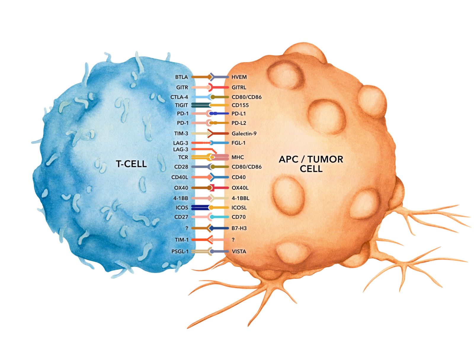
Immune checkpoints are regulators of the immune system which prevent the immune system from attacking self-antigens indiscriminately. While this is critical to preventing auto-immune disease it also causes the immune system to be ineffective in eradicating or suppressing cancer.
Tumor cells exploit certain immune-checkpoint pathways as a major mechanism of immune resistance, particularly against T cells that are specific for tumor antigens. Because many of these immune checkpoints are initiated by ligand-receptor interactions, they can be readily blocked by antibodies. Blocking these immune checkpoints allows antitumor activity to resume and is among the most promising approaches to activating therapeutic anti-tumor immunity.
 Bio X Cell is the main source of in vivo functional antibodies for scientists, offering 300 high quality in vivo pure monoclonal antibodies covering a wide range of your research needs. Bio X Cell InVivoMAb™ in vivo pure monoclonal antibodies are specifically formulated for in vivo use. They feature greater than 95% purity, ultra-low endotoxin levels, and are preservative, stabilizer, and carrier protein-free. The InVivoPlus™ versions of these products are structurally and functionally identical to the InVivoMab™ versions with the added benefit of additional QC measures.
Bio X Cell offers an extensive selection of antibodies targeting mouse immune checkpoint proteins.
Product Highlight: PD-1 (CD279) and PD-L1 (B7-H1)
PD-1 (CD279) is transiently expressed on CD4 and CD8 thymocytes as well as activated T and B lymphocytes and myeloid cells. PD-1 expression declines after successful elimination of antigen. Additionally, Pdcd1 mRNA is expressed in developing B lymphocytes during the pro-B-cell stage. PD-1’s structure includes an ITIM (immunoreceptor tyrosine-based inhibitory motif) suggesting that PD-1 negatively regulates TCR signals. PD-1 signals via binding its two ligands, PD-L1 and PD-L2 both members of the B7 family. Upon ligand binding, PD-1 signaling inhibits T-cell activation, leading to reduced proliferation, cytokine production, and T-cell death. Additionally, PD-1 is known to play key roles in peripheral tolerance and prevention of autoimmune disease in mice as PD-1 knockout animals show dilated cardiomyopathy, splenomegaly, and loss of peripheral tolerance. Induced PD-L1 expression is common in many tumors including squamous cell carcinoma, colon adenocarcinoma, and breast adenocarcinoma. PD-L1 overexpression results in increased resistance of tumor cells to CD8 T cell-mediated lysis. In mouse models of melanoma, tumor growth can be transiently arrested via treatment with antibodies which block the interaction between PD-L1 and its receptor PD-1. For these reasons anti-PD-1 mediated immunotherapies are currently being explored as cancer treatments.
Available anti-mouse PD-1 antibodies from Bio X Cell:
PD-L1 (B7-H1) is expressed on T lymphocytes, B lymphocytes, NK cells, dendritic cells, as well as IFNγ stimulated monocytes, epithelial cells, and endothelial cells. PD-L1 binds to its receptor, PD-1, found on CD4 and CD8 thymocytes as well as activated T and B lymphocytes and myeloid cells. Engagement of PD-L1 with PD-1 leads to inhibition of TCR-mediated T cell proliferation and cytokine production. PD-L1 is thought to play an important role in tumor immune evasion. Induced PD-L1 expression is common in many tumors and results in increased resistance of tumor cells to CD8 T cell-mediated lysis. In mouse models of melanoma, tumor growth can be transiently arrested via treatment with antibodies which block the interaction between PD-L1 and PD-1.
Available anti-mouse PD-L1 antibodies from Bio X Cell:
| Clone 10F.9G2 | InVivoMAb anti-mouse PD-L1 (B7-H1), Art. Nr. BXC-BE0101 |
| InVivoPlus anti-mouse PD-L1 (B7-H1), Art. Nr. BXC-BP0101 |
Other Available Immune Checkpoint Targets:
More Information on Bio X Cell in vivo functional antibodies:
>> InVivoMAb in vivo Functional Antibodies
>> RecombiMAb Recombinant Monoclonal Antibodies
>> InVivoSIM Biosimilar Monoclonal Antibodies
14.09.2022

Protein Ark SpinMate...
Efficiency in protein purification

Tumor Markers
Recombinant Engineered Antibodies

Antibody-Drug Conjug...
ADC Conjugation Kit from ACROBiosystems

Lipid Nanoparticles
Accelerate your Discovery

Cell Adhesion Molecu...
Key Targets Covered by Biorbyt